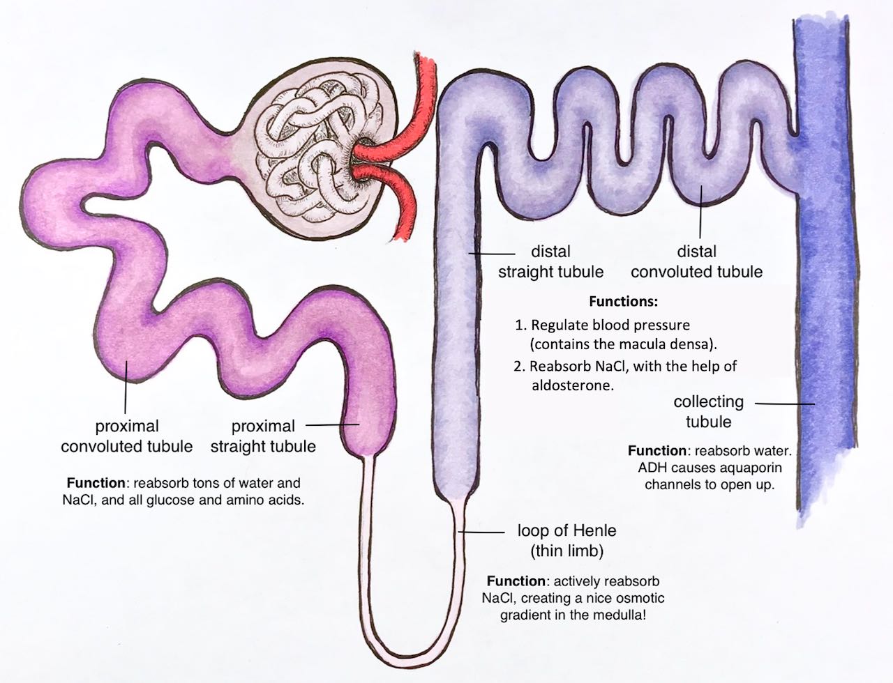
Here’s what happens in each part.
Just a quick note: when we talked about the urinary system in class today, I mentioned that for the last whatever number of years, I thought the macula densa was in the distal straight tubule (that’s what I was taught in medical school, and there are lots of resources that support that). However, when I was preparing for lecture this year, I looked at the literature and it seems like there are even more resources (good ones) that say the macula densa is in the distal convoluted tubule! Perhaps it’s located at the point where the distal straight tubule becomes the distal convoluted tubule. Anyway: the diagram below says the macula densa is in the distal straight tubule, and I just wanted to settle that inconsistency.
Whew. Enough about the macula densa.
Here’s your last challenge: can you remember what each of the segments looks like under the microscope?