Thanks, Beyonce

I can’t help but draw analogies between Beyonce and female reproductive histology. I mean, it’s BEYONCE people. Here she is in all her glory at the 2017 Grammies:

She is adorned in a beautiful golden gown and her head is surrounded by a gorgeous radiating crown. Oh, AND she’s pregnant!

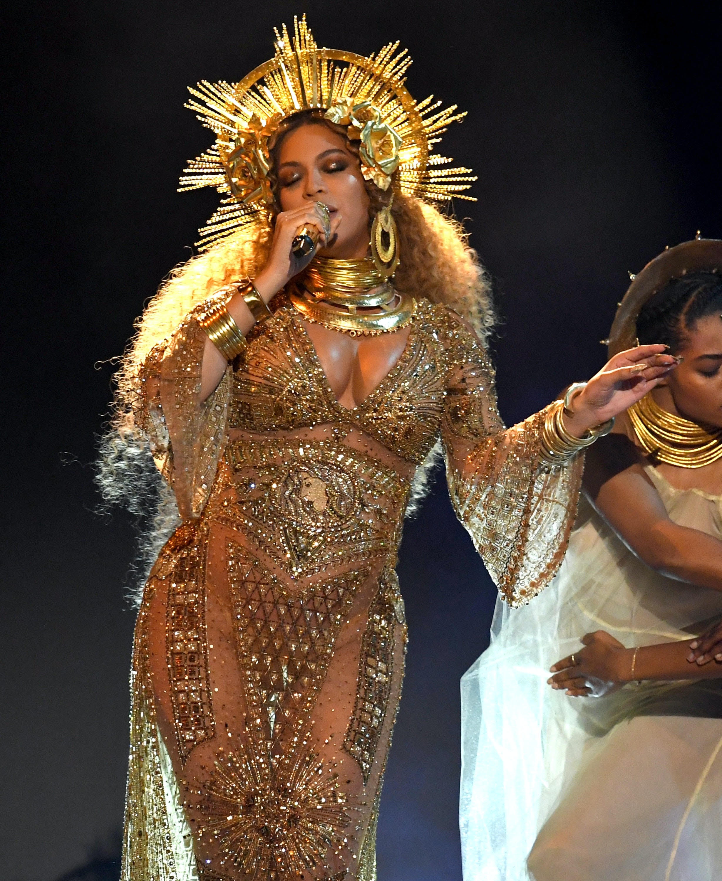
Beyonce is like a primary oocyte sitting in a Graafian follicle: her crown is the corona radiata, and her dress is made of gorgeous granulosa cells. At some point she no doubt sits on a little stool, which is the cumulus oophorus.

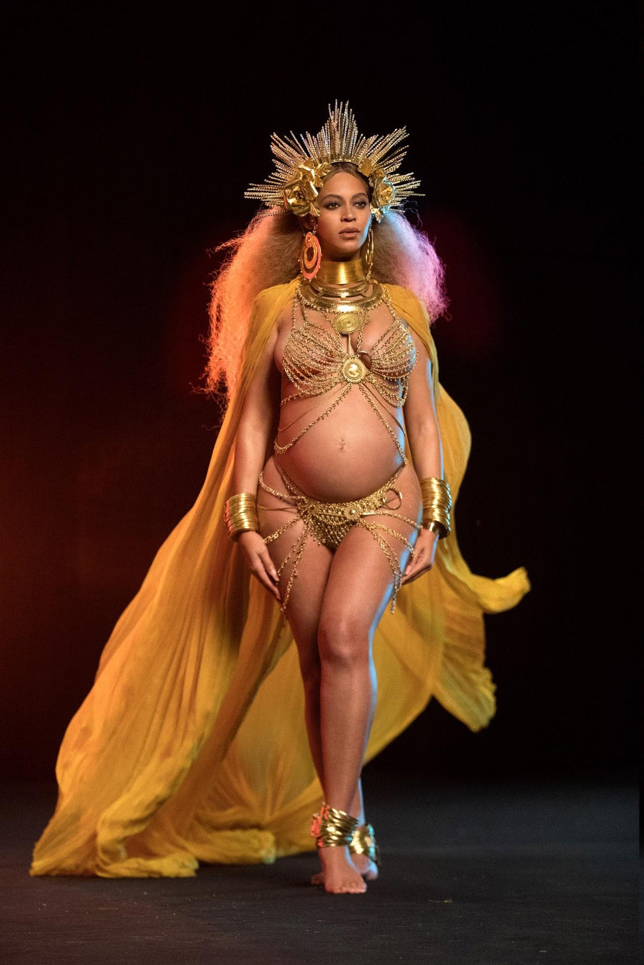
The metaphor continues! What happens during ovulation? Beyonce sheds her gown (the granulosa cells of the Graafian follicle) and leaves the ovary, making her way down that treacherous runway to the opening of the fallopian tube. So what’s left behind? Her gorgeous golden gown! That stays in the ovary, and becomes the corpus luteum (the “yellow body”).

I’m telling you, there’s even more here. If you watch the lecture recording, you’ll see I keep saying the Graafian follicle is juicy (maybe not the best description, but hey, that’s what it looks like to me!). And Bey is most certainly juicy – she straight up says it in Cozy!

Ughhh I love her so much.

Summary of follicle maturation

This diagram shows the different maturational stages of ovarian follicles.


Here’s a quick summary of when you see these different stages:

  1. At birth, all you have are primordial follicles (about a million in each ovary).
  2. At puberty, you start “recruiting” groups of about 50 primordial follicles at the beginning of each menstrual cycle.
    • These primordial follicles start down the pathway of maturation, moving through the stages listed above (unilaminar primary, multilaminar primary, secondary, and Graafian follicles).
    • Only one of the group of 50 follicles will make it to the Graafian follicle stage (the other 49 die off along the way in a process called “attrition” or “atresia”).
    • At the time of ovulation, the Graafian follicle ruptures, releasing the germ cell inside, which makes its way to the fallopian tube and down into the uterus.
  3. In addition, over a woman’s lifetime, the primordial follicles in each ovary start dying off on their own (independent of the menstrual cycles). Their population dwindles over time until the age of menopause, when there are none left. Then menstrual periods cease (there are no primordial follicles left to recruit!).

By the way, here’s what is inside each follicle (spoiler: it’s always a primary oocyte):

  • Primordial follicle: primary oocyte (arrested part way through meiosis I)
  • Unilaminar primordial follicle: primary oocyte (arrested part way through meiosis I)
  • Multilaminar primordial follicle: primary oocyte (arrested part way through meiosis I)
  • Secondary follicle: primary oocyte (arrested part way through meiosis I)
  • Graafian follicle: primary oocyte (arrested part way through meiosis I)

Wait, what?? What about the secondary oocyte and the ovum?

  • At ovulation, the primary oocyte is “released” from its maturational arrest, and it turns into a secondary oocyte. YAY! This secondary oocyte starts undergoing meiosis II, but is arrested part way through.
  • If no fertilization occurs, the secondary oocyte will never complete meiosis II, and will be shed during menstruation.
  • If fertilization occurs, the secondary oocyte completes meiosis II, turning into an ovum. FINALLY!!!

Diagram of what happens to female germ cells

Here’s an interactive drawing I made that shows what happens to female germ cells over time. I think it’s kind of helpful to see the numbers (and stages) of the germ cells mapped out by age – it gives you a visual sense of what happens throughout life.

I can’t post the drawing on our website because of wordpress limitations – but I was able to post it on my Pathology Student website (so when you click on the link, you’ll wind up on that website).